
Disclaimer: machine translated by DeepL which may contain errors.
The Rigakubu News
The Rigakubu News, May 2025.
Research Student Communicates to Faculty >
How Fish Sense Light Directly at the Pituitary Gland
Ayaka Fukuda (Department of Biological Sciences, 3rd Year Doctoral Student) *At the time of research
Shinji Kanda (Atmosphere and Ocean Research Institute / Department of Biological Sciences (concurrent position) Associate Professor)
Have you ever looked closely at the head of a fish?
If you look closely, you will notice that the skull is so transparent that you can faintly see the shape of the brain.
In other words, light reaches the brain. We vertebrates obtain light information from the environment through photoreceptor molecules (opsins) expressed in the eyes
and many non-visual opsins are also expressed in the brain
. However, the physiological significance of these molecules has not been well elucidated.
It is possible that organisms with transparent heads have more light reaching their brains and other parts of their bodies than we humans do, and that they obtain optical information directly from sources other than their eyes.
One such finding was made in the pituitary gland, a hormone-secreting organ located deeper than the brain.
![]()
The pituitary gland is an organ that secretes hormones under the direction of the brain (hypothalamus) and contains cells that secrete various hormones such as gonadotropic hormone, growth hormone, and prolactin. In an experiment to investigate the mechanism of hormone secretion in various cells constituting the pituitary gland, they found that the cells that secrete the hormone that makes the body color darken (melanocyte-stimulating hormone, MSH) (MSH-producing cells) release the hormone in response to light.
The key to this discovery was a technique called Ca2+ imaging. In neurons and hormone-secreting cells (endocrine cells), an increase in intracellular calcium ion concentration ([ Ca2+]i) triggers the release of neurotransmitters and hormones, and we created a genetically modified killifish expressing a Ca2+ indicator (GCaMP) whose fluorescence intensity increases as [ Ca2+ ]i rises. To observe GCaMP, it is necessary to irradiate relatively strong blue light as excitation light. When we irradiated excitation light and observed fluorescence, we noticed that [ Ca2+]i increased. This phenomenon strongly suggested that MSH-producing cells were releasing the hormone in response to light. Detailed examination of this phenomenon revealed that MSH-producing cells express Opn5m, a non-visual opsin that responds most strongly to ultraviolet light (UV-A), and that when Opn5m is activated by short-wavelength light, it raises [ Ca2+]i i via an intracellular signaling pathway (Gq protein and IP3). Therefore, we generated medaka with a knockout of the opn5m gene. These killifish showed no optical response to Ca2+ imaging, indicating that Opn5m is essential for this phenomenon. Next, we used the knockout killifish to investigate what was happening at the individual level: MSH is a hormone that promotes melanin production, and when reared under light conditions including UV-A, gene expression of enzymes involved in epidermal melanin production was lower in the knockout medaka. In addition, in order to analyze the actual blackness of the body color, we quantified the degree of light penetration through the body, and found that light penetration was increased in the knockout killifish. This means that Opn5m on MSH-producing cells is activated by UV light, resulting in the release of MSH and the enhancement of melanin production on the body surface.
These experiments provide a new mechanism by which killifish enhance cellular defense against damaging UV radiation by sensing light directly in the pituitary gland, which is deeper than the brain. It seems that organisms have many more hidden mechanisms that are beyond our intuition.
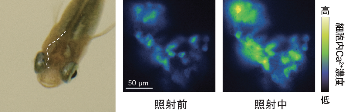 (Left) The head of a killifish is so transparent that the brain can be seen through it. (Left) The head of a killifish is so transparent that the brain can be seen through it. (Middle, right) MSH-producing cells show a clear light response when irradiated by a lamp set at a lower intensity than the light reaching the water.
The results of this study were published in Science, 387,43 (2025) by A. Fukuda et al.
(Press release on January 7, 2025)