DATE2024.09.20 #Press Releases
Discovery of the Mechanism that First Creates the “Inside and Outside” of Plants
—Cells determine the direction of division by slightly changing their shape—
Summary
The basic structure of a plant is cylindrical, like a stem or root. Having a planar internal and external axis connecting the inner vascular bundles to the outer epidermis is important for plant shaping. However, when and how the inner and outer axes are formed has long been a mystery.
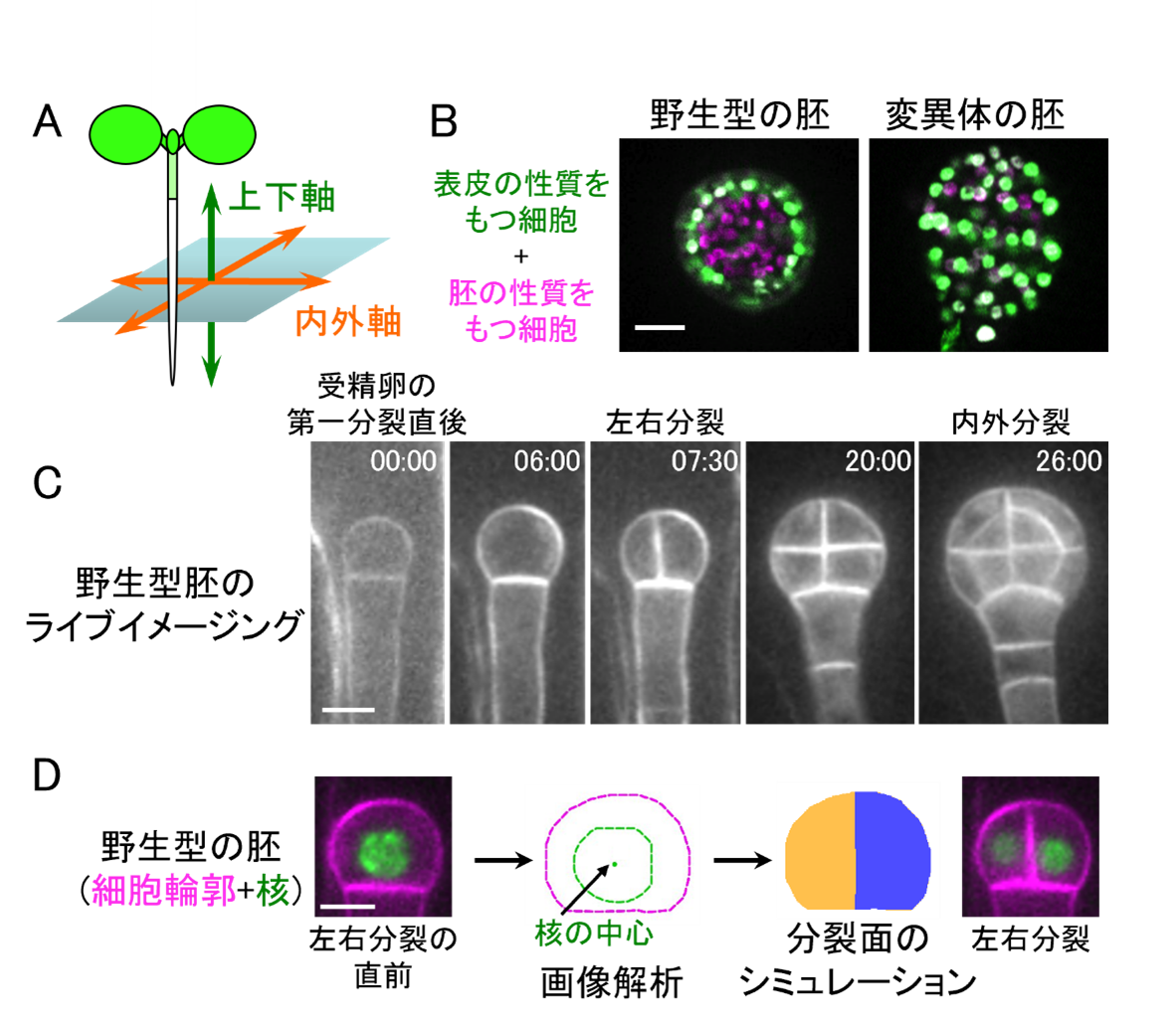
A joint research group led by Professor Minako Ueda of Tohoku University, Professor Koichi Fujimoto of Hiroshima University, Professor Takumi Higaki of Kumamoto University, and Professor Tetsuya Higashiyama of the University of Tokyo has discovered that in the model plant Arabidopsis thaliana, when the HD-ZIP IV transcription factors working in the outermost layers of the embryo are destroyed, the internal and external axes are not well formed. They also found that immediately after the first division of the fertilized egg, the transcription factors cause the cell to elongate slightly horizontally and the nucleus is positioned at the bottom of the cell, and that the cell subsequently divides to the left and right, leading to internal and external divisions. In addition, the cell shape and the nucleus were also found to be related to the cell shape. Furthermore, he found that the cell shape and the position of the nucleus create a plane of division at the mathematically most stable location.
This research revealed an elaborate strategy whereby only small changes in the geometrical information of cell shape and nuclear position are required to determine the direction of division. This discovery is expected to advance our understanding of plant somatic axis formation. The research results were published in Current Biology on September 19, 2024.

Figure:(A) Schematic diagram of a plant body axis. (B) Fluorescently labeled Arabidopsis embryos for the expression of genes working in the epidermis (green) and throughout the embryo (pink). The wild type and the hdg11/12 pdf2 triple mutant, in which the HD-ZIP IV transcription factor cluster is broken, are shown. (C) Live imaging image of a wild-type embryo with fluorescent labeling of the cell membrane. Time indicates hours:minutes. (D) Computer simulation to estimate the direction of division. Based on the cell shape and nuclear position of the wild-type embryo just before left-right division, the “plane of division that passes through the center of the nucleus and has the smallest area” was calculated, showing that the embryo divides left-right as in the actual plane of division. Scale bar represents 10 micrometers (µm).
Professor Tetsuya Higashiyama of the Department of Biological Sciences participated in this research result.
Iinks:Tohoku University, Tohoku University Graduate School of Life Sciences, Hiroshima University, Kumamoto University(in Japanese)
Journal
-
Journal name Current BiologyTitle of paper


