
Disclaimer: machine translated by DeepL which may contain errors.
~ Message from a graduate student~.
Life Science as Seen in Droplets
 |
| Yuta Nakagawa |
| Department of Chemistry, 3rd Year Doctoral Student |
| Birthplace Tokyo, Japan |
| High School Walt Whitman High School (USA) |
| Faculty Skidmore College Department of Chemistry (USA) |
In biology and medicine, the importance of rare cells, which exist only in a very small fraction of the cell population, has been attracting attention in recent years. For example, recent studies have shown that a minority of slow-growing cells with characteristics that could have a major impact on human society, such as resistance to antibiotics and anticancer drugs, are hidden in cell populations. Detailed analysis of these cells is expected not only to provide new insights into basic science, but also to lead to the development of therapeutic drugs and other applications. On the other hand, such rare cells tend to be overlooked by conventional analysis methods, and their detailed ecology and molecular mechanisms are often unknown. Therefore, it is a major challenge for life science to develop an efficient method to search for and isolate rare cells.
In order to find and analyze such rare cells, one-cell analysis is required to measure each cell in a population comprehensively, instead of measuring each cell in a population. In my research, I am utilizing a one-cell analysis technique called droplet microfl uidics. I have developed this technique so that single cells can be encapsulated and cultured in microdroplets of about 100 µm in diameter to search for desired cells. Since dividing cells stay in the same space in the droplet, it is possible to identify cells based on biologically useful indicators such as the speed of cell proliferation, as shown in Figure A. Furthermore, I have developed a method for culturing a large number of cells in a droplet. Furthermore, we developed a device to fractionate only those droplets containing the desired cells from a large population of cell-encapsulated droplets (Figure B). As a result, we succeeded in separating droplets containing budding yeast at a rate of about 1,700 cells per second, realizing a large-scale single-cell analysis using droplets. The speed of preparative separation is important for exhaustive measurement to find rare cells among a large number of cells, and this achievement is expected to be very useful in the search for rare cells. Currently, we are conducting research with the aim of developing this technique into applied research, such as the isolation of drug-resistant cancer cells and the analysis of microglial cells, which are thought to contribute to the progression of Alzheimer's disease.
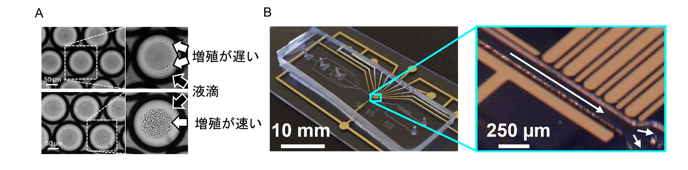
A. After culturing cells in a droplet, slow-growing cells (upper photo) and fast-growing cells (lower photo) can be discriminated. Droplets flow through the channel as indicated by the arrow in the magnified image (right photo). Electrodes placed across the channel generate a local electric field to separate the desired droplets.
The above study is an example of a new method for isolating rare cells being developed in the Structural Chemistry Laboratory of the Department of Chemistry, Graduate School of Science, to which I belong. In our laboratory, graduate students and faculty members from diverse backgrounds conduct interdisciplinary research by bringing together knowledge from various fields. When I visited our laboratory during the summer vacation of my third year as a Faculty member, I was fascinated by the research stance of finding new findings using state-of-the-art technologies developed by our laboratory, and I am still continuing my research. I hope that as many of you as possible will read this article and pursue a career in science together with me.