DATE2022.12.21 #Press Releases
Microbial 'Jenga' : how bacteria stack
December 21, 2022

Figure 1 : A simplified sketch of the attraction of bacterial cells toward topological defects, resulting in a slight increase in height compared to other areas. Illustration credit: Tomo Narashima.
To study the colony's three-dimensional structure, the researchers placed immotile E. coli between a nutrient layer and a thin glass. They observed the bacterial cells multiply and form three-dimensional structures by using confocal and phase-contrast microscopes. To their surprise, the cells stacked vertically in a way that they did not expect.
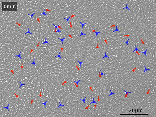
In a nematic system, the constituent particles—in this case, rod-shaped bacteria—are aligned with each other. Examples of a nematic system include bacterial colonies and some filamentous molecules within cells. The points of local mismatches of constituent particle alignments in a nematic are called topological defects. The winding direction of the constituent particles describes the topological defects. For example, Figure 2 shows +1/2 and -1/2 defects in a colony of rod-shaped bacteria.

Figure 2 : Topological defects. The red comet shows a +1/2 defect, and the blue trefoil shows a -1/2 defect. (A) The defects in the bottom layer of a live bacterial colony as viewed through a confocal microscope. (B) Schematics of the defects.
Compared to other locations, the rod cells assembled on top of each other more at the topological defects. And the neighboring cells moved toward both types of defects implying upward growth in those areas. But the conventional active nematic theory suggests that the cells should escape from the -1/2 defects. Dr. Shimaya and Prof. Takeuchi explain this conflicting experimental result using theory. They argue that the three-dimensional tilting of cells on the substrate could add force around the defects pushing the cells toward the -1/2 defects. So, cells move toward the defects and stack on top of each other. To account for their novel findings, Dr. Shimaya and Prof. Takeuchi extended the active nematic theory.

Video 1. Bacterial cells multiply in a colony. As the population density increases, they form three-dimensional mounds.

Video 2. Locations of topological defects and how the cells get tilted. The red comets show +1/2 defects, and the blue trefoils show -1/2 defects.
“So far, we have linked the cell orientation only in the bottom layer (hence two-dimensional) to the initial stage of the three-dimensional colony growth. We are also interested in the three-dimensional structure of cell orientation, which is yet to be characterized but can be relevant to a later stage of colony growth,” says Kazumasa Takeuchi, an Associate Professor at the University of Tokyo.
“In recent years, a series of reports showed how orientation patterns of cell populations influence the way cells coordinate and form supracellular structures. By accumulating experimental facts and refining theory, this research direction may hint at a physical principle that orchestrates living cells. On the application side, it may give, for example, a design principle for suppressing biofilm formation on devices.”
Publication details
Journal PNAS NexusTitle Tilt-induced polar order and topological defects in growing bacterial populationsAuthors DOI


