
DATE2022.11.08 #Press Releases
Discovery of Brain Factors Important for Maintenance of Testicular Morphology and Function in Medaka
Disclaimer: machine translated by DeepL which may contain errors.
Souma Tomihara (Doctoral Student, Department of Biological Sciences at the time of research / currently JSPS Research Fellow)
Hanana Ikegami (At the time of research: JSPS Research Fellow, Department of Biological Sciences / currently Research Fellow, Japan Society for the Promotion of Science)
Graduate School of Agricultural and Life Sciences, The University of Tokyo)
Butiko Shimomai (at the time of research: Master's Program, Department of Biological Sciences)
Chie Matani (Assistant Professor, Department of Biological Sciences at the time of the research, now Assistant Professor, Graduate School of Agricultural Science, Tokyo University of Agriculture and Technology)
Key points of the presentation
- We found that male medaka fish that have lost the function of neuropeptides in the brain have smaller testes after sexual maturity and gradually have difficulty in leaving the next generation.
- NPFF, a type of neuropeptide released from NPFF-producing neurons in the brain, is known to enhance the expression of the follicle-stimulating hormone (FSH) gene, which is thought to be important for testicular development and spermatogenesis in fish, via neurons in the preoptic area.
- This discovery is expected to advance our understanding of the mechanisms in the brain that control the morphology and function of fish testes, which are still poorly understood, and to lead to research aimed at improving methods for aquaculture.
Summary of Presentation
In vertebrate males, it is generally believed that gonadotropic hormone released from the pituitary gland into the bloodstream acts on the testes to regulate their development and maintenance of function. This mechanism has been well studied in mammals, and specific neural circuits in the brain that regulate the production and release of gonadotropic hormone have been identified. In vertebrates other than mammals, such as echinoderms, however, many of the neural circuits in the brain that regulate these functions remain unknown.
A research group at the Graduate School of Science, The University of Tokyo, conducted a detailed analysis of the morphology and function of the testes of male medaka fish in which the function of neuropeptide FF (NPFF) (Note 1), a type of neuropeptide expressed in the brain, was lost using genome editing technology. As a result, we found that the testes of male medaka lacking NPFF function regress after sexual maturity, and it gradually becomes difficult for the medaka to leave a next generation. Further analysis of the neural circuits in the brain revealed that NPFF released from neurons (Note 2) in the region called the terminal nerve acts on neurons in the preoptic area that extend their axons to the pituitary gland, ultimately inducing follicle-stimulating hormone (FSH), a type of gonadotropic hormone that is important for testicular function in mammals. (FSH) (Note 3), a type of gonadotropic hormone important for testicular function in mammals. This discovery is expected to advance our understanding of the mechanisms in the brain that regulate testicular development and function in male fish.
The research results will be published online in The Proceedings of the National Academy of Sciences of the United States of America the week of November 7, 2022 (EST).
Announcement
Most vertebrates have two sexes, female and male, with the female producing eggs in the ovaries and the male producing sperm in the testes. The development and maintenance of ovarian and testicular function are thought to be regulated by specific neural circuits in the brain and hormone secretion controlled by these circuits. In mammals, it is known that gonadotropin-releasing hormone (GnRH)-releasing neurons in the brain regulate the release of gonadotropin in the pituitary gland, resulting in ovarian and testicular development. However, many mysteries remain regarding the mechanisms in the brain that control the morphology and maintenance of testicular function in vertebrates other than mammals, as it has been reported that male medaka fish with loss of GnRH function have normal reproductive function (Takahashi et al., 2016). The research group focused on NPFF, a type of neuropeptide released by specific neurons in the brain, and conducted an analysis using male killifish that cannot produce NPFF in the brain (medaka with loss of NPFF function).
First, male killifish with loss of NPFF function were paired with normal females, and the fertilization rate of eggs laid by the females was examined. Normal male-female pairs had a fertilization rate of 80-100% at 5 or 6 months of age, but when paired with killifish that had lost NPFF function, the fertilization rate dropped significantly to an average of 40% at the same age, and some pairs produced no fertilized eggs at all (Figure 1 (A)). We then measured the size of the testes of male killifish that had lost NPFF function and found that they were significantly smaller than those of normal males (Figure 1(B)). Next, histological analysis of the testes of the NPFF loss-of-function male medaka revealed that, despite their smaller size, normal testicular structures were produced and spermatogenesis was taking place. This suggests that NPFF produced in the brain works in some way to maintain the testes as large as they are.
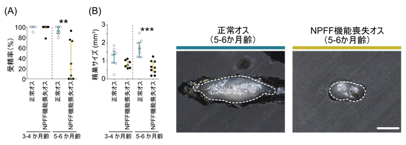
Figure 1: Fertilization rate and morphology of male medaka with loss of NPFF function.
(A) Normal males and males with loss of NPFF function were mated with normal females, and the fertilization rate of eggs laid by the females was measured. The fertilization rate of normal females paired with 5- to 6-month-old males with loss of NPFF function was significantly lower. (B) The testes of NPFF loss-of-function males were significantly smaller than those of normal males. Scale bar: 1 mm
Therefore, we measured blood levels of 11-ketotestosterone (11-KT), a type of male steroid hormone that is believed to be involved in maintaining testicular function in fish, and found that the levels were significantly lower in 5- and 6-month-old NPFF loss-of-function male medaka. On the other hand, when the expression of the gene required for 11-KT biosynthesis was quantified in the testes, its expression was higher in the NPFF loss-of-function male medaka, in contrast to the lower concentration of 11-KT. Further studies on how changes in 11-KT concentration are related to the maintenance of testis size are warranted.
Next, we investigated the pathway by which NPFF produced in the brain acts on the testes. First, to determine whether NPFF acts directly on the testes, we checked whether NPFF receptors are expressed in the testes, and found that none of the three types of NPFF receptors are expressed. In response, we analyzed the gene expression of follicle-stimulating hormone (FSH), a type of gonadotropic hormone produced in the pituitary gland that is thought to enhance the synthesis of steroid hormones in the fish testis. The results showed that FSH expression was significantly lower in NPFF loss-of-function male medaka. Furthermore, among the neurons in the brain that extend axons to the pituitary gland, neurons in a region called the preoptic area were found to express receptors for NPFF. Previous studies have shown that the preoptic area is the region where neurons in the terminal nerve that release NPFF extend their axons ( Oka and Matsushima, 1993; Umatani et al., 2022). These findings suggest that in male fish, NPFF released from neurons of the terminal nerve in the brain is received by NPFF receptor-expressing neurons in the preoptic area, and through some signal released by these neurons, the expression of FSH in the pituitary gland is enhanced, thereby maintaining the morphology and function of the testis This finding suggests that FSH expression in the pituitary gland is upregulated by some signal released from these neurons (Fig. 2). This discovery is expected to advance our understanding of the mechanisms in the brain that govern the development and maintenance of fish testis function, which is still poorly understood, and to lead to research for the improvement of aquaculture methods.
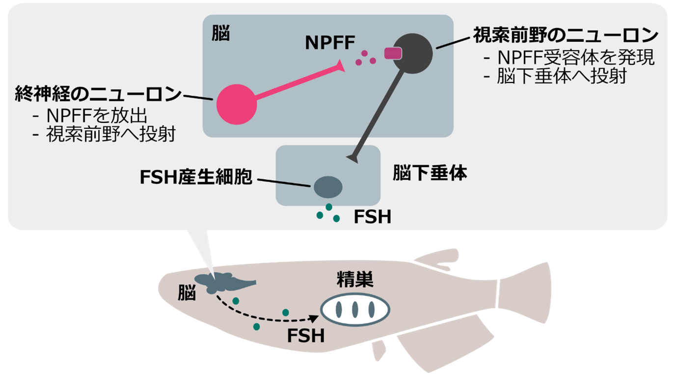
Figure 2: Working hypothesis on how NPFF maintains testicular morphology and function.
NPFF released from neurons in the terminal nerve is received by neurons whose cell bodies are localized in the preoptic area and express NPFF receptors. These neurons extend their axons to the pituitary gland, suggesting that they somehow enhance the expression of the FSH gene in pituitary FSH-producing cells. The FSH produced by this mechanism reaches the testes via the blood circulation and may be involved in the maintenance of testicular morphology and function.
This research was supported by Grant-in-Aid for Scientific Research (Grant-in-Aid for Scientific Research No. 20H03071 and 22K19326) and Grant-in-Aid for Young Scientists (Grant-in-Aid for Young Scientists No. 19J21828, 22J01130, and 19J00450).
(References)
Takahashi et al., Endocrinology, 157 (10):3994-4002, 2016
Oka and Matsushima, Journal of Neuroscience, 13 (5): 2161-2176, 1993
Umatani et al. , Endocrinology, 163 (2), bqab261. 2022
Journal
-
Journal name The Proceedings of the National Academy of Sciences of the United States of America Title of paper Neuropeptide FF indirectly affects testicular morphogenesis and functions in medaka Author(s) Soma Tomihara, Kana Ikegami, Rinko Shimomai, and Chie Umatani* (University of California, Berkeley) DOI Number
Terminology
1 Neuropeptide FF (NPFF )
A type of peptide called RF amide with a characteristic amino acid sequence at the C-terminal side, which is produced by neurons in the region of the brain known as the terminal nerve. It was known to be involved in the sexual behavior of fish males, but whether it is involved in the maintenance of gonadal function has not yet been clarified. ↑up
Note 2 Neuron
Synonymous with neuron. Neurons have many neurites, which release neuropeptides and other chemical substances, and exchange signals among neurons to produce sensory, motor, and other brain functions. ↑up
Note 3 Follicle-stimulating hormone (FSH )
A type of gonad-stimulating hormone secreted by the pituitary gland that causes gonadal development, etc. via systemic blood circulation. In fish, it is thought to enhance the synthesis of steroid hormones in the testes. ↑up