
DATE2022.11.04 #Press Releases
Elucidate the whole picture and differences between individual mother-derived cell types that are transferred during fetal life.
Disclaimer: machine translated by DeepL which may contain errors.
Kana Fujimoto (Department of Biological Sciences, Doctoral Program at the time of research)
Yumiko Tanaka, Doctoral Student (at the time of research), Department of Biological Sciences
Soutarou Uemura, Professor, Department of Biological Sciences
Naoki Irie, Associate Professor, Department of Biological Sciences
Key points of the presentation
- We have revealed the cell types of mother-derived cells that are transferred into mouse fetuses during pregnancy, and have shown that the proportions of immune-related cells and stem cell-like cells vary greatly from fetus to fetus.
- Although various types of mother-derived cells have been reported in humans, it was not clear whether the cell types of mother-derived cells differ from fetus to fetus (individual) or not.
- Future research focusing on inter-individual differences in mother-derived cell types will contribute to the elucidation of the causes of various in vivo phenomena that are thought to be related to mother-derived cell types.
Summary of Presentation
Associate Professor Naoki Irie and Kana Fujimoto (then a graduate student) of the Graduate School of Science, The University of Tokyo, and their colleagues have elucidated the full picture of cell types of mother-derived cells collected from each individual mouse fetus, based on the gene expression information of each cell.
Mammals with placentas (placental mammals), including humans and mice, are believed to transfer their mother's cells to the fetus when it is in the mother's womb, and these cells remain in the fetus throughout its life. These maternal cells are called "mother-derived cells" (Figure 1). Previous studies in our laboratory have shown that the number of mother-derived cells differs among individuals, but this study has revealed that the types (repertoire) of mother-derived cells also differ among individuals. Mother-derived cells have been suggested to be involved in various biological phenomena, such as avoidance of immunological conflicts between mother and offspring, regeneration of offspring tissues, and onset and exacerbation of diseases, but it is unclear why they can be involved in such diverse phenomena. This research is expected to provide important fundamental knowledge in elucidating this mystery.
The research results were published online in the international scientific journal Scientific Reports on Friday, November 4, 2022, at 7:00 p.m.
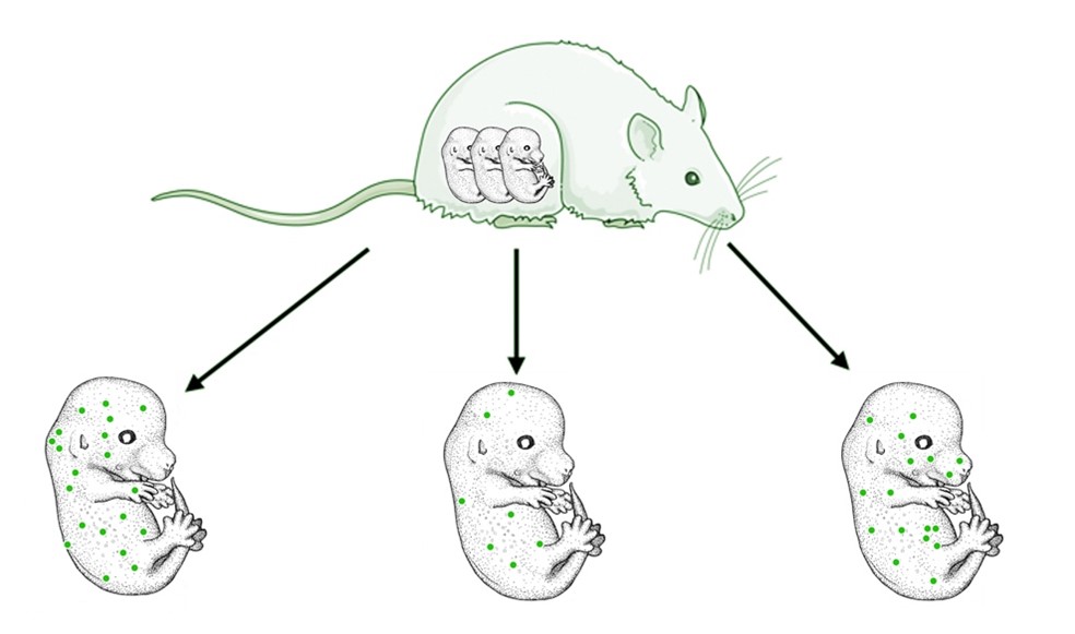
Figure 1: Mother-derived cells are transferred during fetal life and remain viable in the child after birth.
Mammals with placentas (placental mammals), including humans and mice, transfer cells from the mother to the fetus when it is in utero, and these cells remain in the fetus for the rest of its life. These maternal cells are called "mother-derived cells. In this study, we found that not only the number of mother-derived cells but also the cell type differs among fetuses (individuals). In the figure, mother-derived cells are indicated by green dots.
Publication
In placental mammals, including humans, cells are transferred from the mother to the fetus during pregnancy, and these mother-derived cells remain in the fetus throughout its entire body, even after birth, although in minute amounts (1:100,000 to 1:10 million frequency). It has been suggested that mother-derived cells are involved in a variety of phenomena, such as avoidance of immune system conflicts between mother and child, onset and exacerbation of inflammatory diseases in the offspring, and repair of damaged tissues. However, it remains unclear why all individuals undergo transfer of mother-derived cells, yet different individuals have been shown to be involved in different phenomena, and the mechanisms that generate these differences remain unclear.
The research group hypothesized that one possible explanation for these differences could be individual differences in the number and type of mother-derived cells transferred during fetal life. In fact, a high frequency of mother-derived cells (10 to 100 times higher than in healthy subjects in some tissues) has been observed in inflammatory diseases, and a number of mother-derived cells of various cell types, such as immunocompetent cells and stem cells, have been reported in each phenomenon. However, due to the low frequency of mother-derived cells, no previous studies have comprehensively examined whether the number and types of mother-derived cells differ among fetuses (or individuals). Clarification of this question is considered to be fundamental research for verifying the causal relationship between mother-derived cells and various phenomena that are thought to be related to mother-derived cells, and is also important for understanding the biological significance of mother-derived cells.
In this study, we used mice, which are representative laboratory animals of placental mammals, to test the above hypothesis. Although our previous study (Fujimoto et al. PLOS ONE (2021) 16(12): e0261357) showed that there are individual differences in the number of mother-derived cells throughout the mouse fetus, there has been no comprehensive study that comprehensively examined whether cell types differ from fetus to fetus Therefore, the present study was designed to investigate whether the number of cells derived from the mouse fetus differs between fetuses. Therefore, in this study, we determined the cell type of the mother-derived cell population in each individual mouse fetus and examined whether there are inter-individual differences in the mother-derived cell type.
First, we labeled mother-derived cells with fluorescent protein (GFP) and constructed an experimental system to detect only GFP-positive cells from whole mouse fetuses (E14.5) in the early stages of mother-derived cell transfer. To minimize the loss of mother-derived cells, which are few in number, we isolated mother-derived cells (GFP-positive cells) by flow cytometry (FACS) (Note 1) for each individual fetus, using a fetal tissue isolation system in which cell death was as unlikely as possible. Next, gene expression data were obtained for each isolated cell (single cell RNA-seq) (Note 2), and the cell type of the mother-derived cells was estimated and analyzed by comparing the data with the known gene expression information database for each cell type (Note 3), and then confirming the gene information expressed by each mother-derived cell. The results showed that the analyzed embryonic mouse embryos were of the same type as the embryonic mouse embryos.
As a result, we were able to isolate 191 GFP-positive cells from 26 of the 52 mouse fetuses analyzed, which were considered to be of maternal origin. Averaging the cell types inferred from the gene expression patterns of each cell, it was found that approximately 67% of the mother-derived cells were immunocompetent cells, 25% were terminally differentiated (Note 4), and the remaining 8% were stem cell-like cells with the ability to divide (Figure 2A, B). Furthermore, when examined on an individual basis, the proportion of immunocompetent cells and stem cell-like cells was found to vary widely (Figure 3). This is the first result showing that the cell types of mother-derived cells differ among individuals (fetuses).
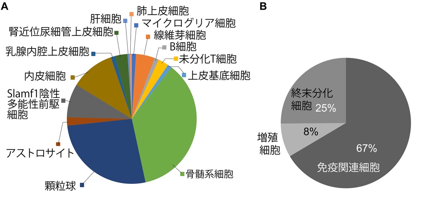
Figure 2: Estimated cell types of mother-derived cells and their proportions (all individuals overall)
A: Average percentage of cell types estimated from gene expression patterns of 191 GFP-positive cells isolated from 26 mouse fetuses.
B: Based on the cell type estimation in A, the results were converted into the percentages of immunocompetent cells, mitotically competent stem cell-like cells, and terminally differentiated cells.
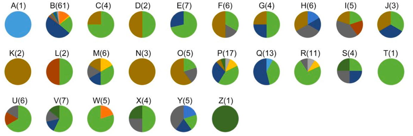
Figure 3: Percentage of estimated mother-derived cell types in each individual.
The estimated cell types and their percentages are shown for each mouse fetus from which mother-derived cells were isolated. Alphabets above the pie chart label the individual, and the number of cells isolated from that individual is listed in parentheses. Each color in the graph corresponds to Figure 2A.
We also found a feature common to many mother-derived cells. When we examined the genes commonly expressed in mother-derived cells, we found that the gene encoding a protein that may be related to cell migration ( Interferon-induced transmembrane protein 2 (Ifitm2) gene) is commonly expressed in more than 90% of mother-derived cells The IGF-1 gene, which belongs to the same family as this protein, is expressed in more than 90% of mother-derived cells. Ifitm3 gene, which belongs to the same family as this protein, is known to be expressed in migrating primordial germ cells. Expression of the Syndecan-binding protein (Sdcbp) gene, which is involved in cell migration and human tumor metastasis, was also confirmed. Although further studies are needed, mother-derived cells may be migrating to the fetal side using these genes/proteins related to cell migration.
This study revealed that there are individual differences in the mother-derived cell types in mouse fetuses. Just as the genetic information passed from parent to offspring differs slightly from sibling to sibling, the information passed from mother to offspring also differs at the cellular level. In addition, among the in vivo phenomena in which mother-derived cells have been suggested to be involved, many of the causes of the onset and aggravation of autoimmune diseases, especially in the offspring, are unknown. In the future, if the reasons for inter-individual differences in mother-derived cell types can be clarified and the proportion of each cell type in mother-derived cells can be controlled, it may lead to symptomatic improvement and regeneration of inflammatory diseases in which mother-derived cells are thought to be involved, as well as to promotion of immune tolerance.
This research was supported by a Grant-in-Aid for Scientific Research (Grant-in-Aid for Scientific Research No. 21K19254), the Takeda Foundation for the Promotion of Science, and a Grant-in-Aid for Distinguished Contributors (DC2).
Members of this research team
| Kana Fujimoto | D. Student (at the time of research), Department of Biological Sciences, Graduate School of Science, The University of Tokyo |
| Kei Nakajima | Assistant Professor, Department of Pharmacy, Graduate School of Pharmaceutical Sciences, The University of Tokyo |
| Shohei Hori | Professor, Department of Pharmacy, Graduate School of Pharmaceutical Sciences, The University of Tokyo |
| Yumiko Tanaka | Doctoral Student (at the time of research), Department of Biological Sciences, Graduate School of Science, The University of Tokyo |
| Yoshitaka Shirasaki | Project Assistant Professor, Graduate School of Pharmaceutical Sciences, The University of Tokyo |
| Sotaro Kamimura | Professor, Department of Biological Sciences, Graduate School of Science, The University of Tokyo |
| Naoki Irie | Associate Professor, Department of Biological Sciences, Graduate School of Science, The University of Tokyo |
Journal
-
Journal name Scientific Reports Title of paper Whole-embryonic identification of maternal microchimeric cell types in mice using single-cell RNA sequencing Author(s) Kana Fujimoto*, Akira Nakajima, Shohei Hori, Yumiko Tanaka, Yoshitaka Shirasaki, Sotaro Uemura, Naoki Irie*, Yoshitaka Shirasaki DOI Number
Terminology
1 Flow cytometry (FACS )
An experimental device that enables information on individual cells to be obtained by passing cells through a liquid and measuring scattered light and fluorescence by shining a laser beam on them, as well as isolating only the cells of interest. ↑up
Note 2 Single cell RNA-seq
A method to examine gene expression in each cell based on the RNA contained in a single cell. ↑up
3 Database of gene expression information by known cell types
Tabula Muris from N. Schaum et al.
Note 4 Terminal differentiation
A cell in which cell specialization (differentiation) has occurred to the end. ↑