
DATE2024.02.29 #Press Releases
Development of Near-Infrared Fluorescent Labeling Agent for Live Cell Imaging with Excellent Light Resistance
Disclaimer: machine translated by DeepL which may contain errors.
~Discovery that cell membrane permeability differs depending on stereochemistry~.
Nagoya University
RIKEN
School of Science, The University of Tokyo
Japan Science and Technology Agency
Summary of Presentation
A research group led by Masayasu Taki, Project Associate Professor, Shigehiro Yamaguchi, Professor, and Florence Tama (Team Leader, RIKEN Center for Computational Science) at the Institute for Transformative Biomolecular Research (WPI-ITbM*), Nagoya University, Professor Yasushi Okada (Team Leader, RIKEN Center for Frontier Biosciences) at the University of Tokyo have successfully developed a near-infrared fluorescent labeling agent for live cell imaging) that combines high light resistance and cell membrane permeability.
Light in the near-infrared region is less toxic to living organisms and less susceptible to autofluorescence, making it suitable for long-term imaging of live cells. It is also useful for multicolor staining of targets because its wavelength range does not overlap with that of widely used fluorescent proteins and fluorescent labeling agents in the visible light range. However, most near-infrared fluorescent dyes have low water solubility and chemical stability and are degraded by light, so their availability for live-cell imaging of organelles, which requires long-term observation, has been very limited.
In this study, we developed a near-infrared fluorescent labeling compound with excellent light stability and evaluated its cell membrane permeability. As a result, we found that stereochemistry, the three-dimensional structure of a compound, has a significant effect on its cell membrane permeability. By using a near-infrared fluorescent labeling agent with membrane permeability, we achieved specific labeling of any organelle by fluorescence imaging. We also succeeded in tracing the interaction between organelles by super-resolution 5D imaging (3D + time + wavelength).
These research results were published in the online edition of the German Chemical Society's journal "Angewandte Chemie International Edition" on February 27, 2024.
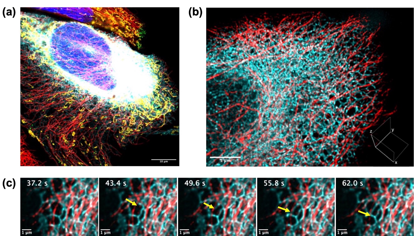
Figure: (a) Multicolor imaging of a living cell organelle. Blue: nucleus, green: lysosome, yellow: mitochondria, red: microtubules, cyan: endoplasmic reticulum (b) Super-resolution 3D imaging of cell structure. Red: microtubule (SiR-Tubulin), cyan: endoplasmic reticulum (trans-1) (c) Network structure of ER changing over time.
For more information, please visit the Nagoya University website.
Journals
-
Journal name Title of paper Stereochemistry-Dependent Labeling of Organelles with a Near-Infrared-Emissive Phosphorus-Bridged Rhodamine Dye in Live-Cell Imaging